


Content Here
Content Here
Presentation Scan
Presentation Scan
More
Scans:

Presentation Scan
![]() Appointment length: 15 minutes.
Appointment length: 15 minutes.
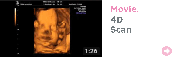
![]() A selection of still 2D scan images.
A selection of still 2D scan images.
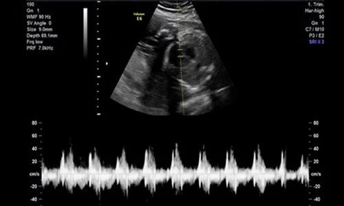
![]() Purpose of the scan: To check the presentation of the baby, head down or breech. We also check the measurements of baby.
Purpose of the scan: To check the presentation of the baby, head down or breech. We also check the measurements of baby.
Optional Add one of our heartbeat bears to this package for £20.


Presentation Scan
A Presentation Scan is performed in order to examine the position of the baby and also the placenta. This type of ultrasound is carried out in the third trimester between 34 – 40 weeks of pregnancy. This is when baby begins to take the “best fit” position for delivery. The ultrasound determines the foetal lie and presentation prior to delivery so that the delivery can be managed in the proper clinical manner.
The ultrasound will check the position of the placenta to check for placenta previa, this is a condition in which the placenta is low in the uterus, next to or covering the cervix. If placenta previa is diagnosed early in pregnancy it may not present a problem in the latter stage of pregnancy as it is likely to migrate; however later in the pregnancy bleeding may occur and complications arising from this may mean that you will have to deliver early by caesarean section.
The presentation refers to the part of the baby presenting at the opening of cervix. The most common and safest for delivery is Cephalic or head down presentation where the head of the baby is in the maternal pelvis. Breech presentation is where the baby is bottom down and the rump is presenting at the opening of the cervix.
In the event that the fetus is in a breech position, a vaginal delivery is made more complicated. Vaginal delivery is the preferred method of delivery as it involves less risk for the mother; therefore, it is possible the obstetrician may want to perform a procedure called External Cephalic Version (ECV.) This is where the fetus is maneuvered using gentle pressure through the mother’s abdomen into the cephalic position (head first.) Medication to relax the abdominal muscles can be used to increase the success of the procedure. ECV can be performed from 36 weeks up to the time of delivery however the earlier it is carried out the better chance of success there is.
You are generally only offered a presentation scan on the NHS if your midwife is not convinced that baby has turned to a head down position, at Baby Scanning you do not need an NHS referral to attend our clinic. This is why Baby Scanning strive to provide a service that is both professional and affordable.
Purpose of the scan
Determine the position of baby and the placenta
What happens during an ultrasound scan?
Our Presentation Scan is available from 34 -40 weeks of pregnancy, your appointment will last approximately 15 minutes, and will be performed on your abdomen. During your appointment the sonographer will ask that you lay on the bed and lift your top up, ultrasound gel will be placed on your abdomen, the sonographer will then make all relevant checks and also let you know the position of baby and the placenta. They will also take some images of your baby which will be available to take home on the day, as this is a later stage in pregnancy and there is not much space and the baby can be quite crammed which makes it difficult to get 4D images, however the sonographer will do their best to get you images.
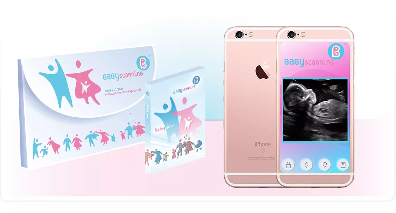
To book your scan please use our online booking system or call our friendly booking line team who will be happy to help. Lines are opened from 9am – 9pm.


Title Here
Content Here
Title Here
Content Here
Title Here
Content Here
Presentation Scan

![]()


More Scans:
Presentation Scan
Presentation Scan
![]() Appointment length: 15 minutes.
Appointment length: 15 minutes.
![]() A selection of still 2D scan images.
A selection of still 2D scan images.
![]() Purpose of the scan: To check the presentation of the baby, head down or breech. We also check the measurements of baby.
Purpose of the scan: To check the presentation of the baby, head down or breech. We also check the measurements of baby.
Optional Add one of our heartbeat bears to this package for £20.
![]()
Presentation Scan
A Presentation Scan is performed in order to examine the position of the baby and also the placenta. This type of ultrasound is carried out in the third trimester between 34 – 40 weeks of pregnancy. This is when baby begins to take the “best fit” position for delivery. The ultrasound determines the foetal lie and presentation prior to delivery so that the delivery can be managed in the proper clinical manner.
The ultrasound will check the position of the placenta to check for placenta previa, this is a condition in which the placenta is low in the uterus, next to or covering the cervix. If placenta previa is diagnosed early in pregnancy it may not present a problem in the latter stage of pregnancy as it is likely to migrate; however later in the pregnancy bleeding may occur and complications arising from this may mean that you will have to deliver early by caesarean section.
The presentation refers to the part of the baby presenting at the opening of cervix. The most common and safest for delivery is Cephalic or head down presentation where the head of the baby is in the maternal pelvis. Breech presentation is where the baby is bottom down and the rump is presenting at the opening of the cervix.
In the event that the fetus is in a breech position, a vaginal delivery is made more complicated. Vaginal delivery is the preferred method of delivery as it involves less risk for the mother; therefore, it is possible the obstetrician may want to perform a procedure called External Cephalic Version (ECV.) This is where the fetus is maneuvered using gentle pressure through the mother’s abdomen into the cephalic position (head first.) Medication to relax the abdominal muscles can be used to increase the success of the procedure. ECV can be performed from 36 weeks up to the time of delivery however the earlier it is carried out the better chance of success there is.
You are generally only offered a presentation scan on the NHS if your midwife is not convinced that baby has turned to a head down position, at Baby Scanning you do not need an NHS referral to attend our clinic. This is why Baby Scanning strive to provide a service that is both professional and affordable.
Purpose of the scan
Determine the position of baby and the placenta
What happens during an ultrasound scan?
Our Presentation Scan is available from 34 -40 weeks of pregnancy, your appointment will last approximately 15 minutes, and will be performed on your abdomen. During your appointment the sonographer will ask that you lay on the bed and lift your top up, ultrasound gel will be placed on your abdomen, the sonographer will then make all relevant checks and also let you know the position of baby and the placenta. They will also take some images of your baby which will be available to take home on the day, as this is a later stage in pregnancy and there is not much space and the baby can be quite crammed which makes it difficult to get 4D images, however the sonographer will do their best to get you images.
To book your scan please use our online booking system or call our friendly booking line team who will be happy to help. Lines are opened from 9am – 9pm.
![]()
![]()